
Revolutionary Neuroimaging Technology Achieves Unprecedented 64 Million-fold Increase in Brain Image Clarity
technology
Cutting-edge advancements in neuroimaging technology have ushered in a remarkable breakthrough - the resolution of brain images has been enhanced to a staggering 64 million times sharper than before.

In a decades long technical tour de force led by Duke’s Center for In Vivo Microscopy with their colleagues at the University of Tennessee Health Science Center, University of Pennsylvania, University of Pittsburgh and Indiana University, researchers took up the gauntlet and improved the resolution of MRI leading to the sharpest images ever captured of a mouse brain.
Coinciding with the 50th anniversary of the first MRI, the researchers generated scans of a mouse brain that are dramatically crisper than a typical clinical MRI for humans, the scientific equivalent of going from a pixelated 8bit graphic to the hyper realistic detail of a painting. One of the major technological advancements that has revolutionized the field of neuroscience is neuroimaging technology. Neuroimaging techniques allow scientists to obtain images of the brain in order to better understand its structure, function and activity.
In recent years, cutting-edge advancements in neuroimaging technology have enabled researchers to achieve an unprecedented level of detail in brain imaging. One such breakthrough is the enhancement of resolution in brain images to a staggering 64 million times sharper than before. This has allowed scientists to see the brain in greater detail than ever before, down to the level of individual neurons and their connections.

One of the key neuroimaging techniques that has enabled this breakthrough is called magnetic resonance imaging (MRI). MRI uses powerful magnetic fields and radio waves to produce detailed images of the brain. Advances in MRI technology have allowed scientists to improve the resolution of these images, making it possible to see the brain at a level of detail that was once unimaginable.
Another important neuroimaging technique that has contributed to this breakthrough is called functional magnetic resonance imaging (fMRI). fMRI measures changes in blood flow in the brain in response to different stimuli, allowing researchers to map out areas of the brain that are active during particular tasks or experiences. Advances in fMRI technology have also contributed to the enhanced resolution of brain images.
The Role of MRI and fMRI in Achieving Unprecedented Clarity of Brain Images
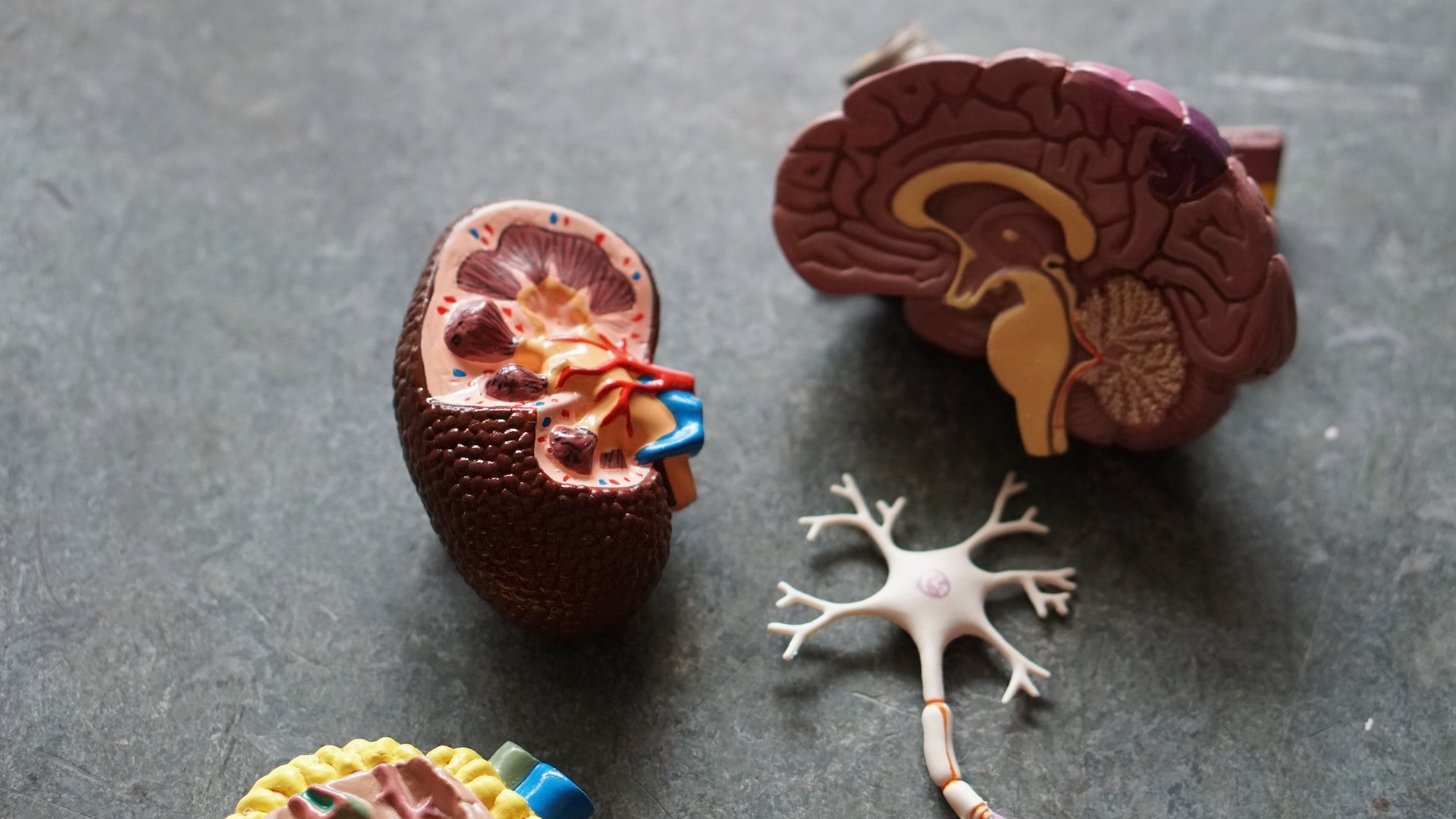
Magnetic Resonance Imaging (MRI) is an advanced technology that allows us to capture vivid images of soft, aqueous tissue that is difficult to visualize using X-rays. However, despite its proficiency in detecting brain tumors, MRI's ability to reveal microscopic details of the brain's structure has been a point of concern.
Nevertheless, the Duke Center for In Vivo Microscopy, in collaboration with prestigious institutions such as the University of Pennsylvania and the University of Pittsburgh, embarked on a technical feat that lasted for decades. Their aim was to enhance the resolution of MRI to capture the minutest details of the mouse brain. Their hard work paid off, as they managed to generate images of the mouse brain that surpass any clinical MRI intended for human use.

Horizontal ‘slices’ of the circuitry data moving up and down across the brain
Despite the use of mice as subjects for the experiment, the researchers have developed a highly sophisticated MRI technique that allows for unprecedented visualization of the entire brain's connectivity. This breakthrough will undoubtedly pave the way for novel insights into the effects of various factors on brain function in humans, such as the impact of aging, dietary habits and neurodegenerative conditions like Alzheimer's disease.
By leveraging the power of mouse imaging, the researchers have opened up a promising avenue for unraveling the mysteries of the human brain and developing innovative treatments for brain related disorders.
Brain Connectivity: Advancing MRI Resolution through Mouse Imaging for Groundbreaking Insights and Innovative Treatments
After almost 40 years of research, the Duke's Center team have achieved groundbreaking MRI resolution for In Vivo Microscopy. Their work, featured in the Proceedings of the National Academy of Sciences on April 17, combines several key elements including a 9.4 Tesla magnet, gradient coils 100 times stronger than clinical MRIs, and a high performance computer equivalent to nearly 800 laptops all working together to image the brain.
The team scans tissues thoroughly and uses light sheet microscopy to label specific groups of cells, such as dopamine issuing cells, to observe Parkinson's disease progression. They then combine the highly accurate light sheet pictures with the anatomically accurate MRI scan, allowing researchers to examine microscopic details of the brain in unprecedented ways. This approach reveals changes in brain wide connectivity as mice age and the deterioration of neural networks in Alzheimer's disease.
By improving the MRI's power as a microscope, researchers hope to gain a better understanding of human diseases such as Alzheimer's and Huntington's. Researchers plans to investigate whether animals on modest dietary and drug interventions, which can extend their lifespan by 25%, maintain cognitive function, providing insights into the human condition.
The researchers team employ light sheet microscopy to label and track specific groups of cells, including dopamine issuing cells in Parkinson's disease. By combining this data with highly detailed MRI scans, the researchers gain unprecedented insight into brain cells and circuits.
This approach reveals brain wide connectivity changes as mice age and showcases the deterioration of neural networks in a mouse model of Alzheimer's disease. By using MRI as a high-powered microscope, researchers hope to better understand human diseases and potential interventions. This research has the potential to uncover whether modest interventions can lead to extended lifespan with an intact brain.
The Bottom Line
The revolutionary neuroimaging technology that has achieved an unprecedented 64 million-fold increase in brain image clarity is truly remarkable. The implications of this breakthrough are vast and far-reaching, and the potential benefits for medical diagnosis and treatment are enormous.
As this technology continues to advance and be refined, we can only imagine the further possibilities that may emerge. With such incredible clarity in brain imaging, researchers and clinicians will be able to gain new insights into the brain's workings and find more effective ways to treat neurological disorders.
It is exciting to see such progress in the field of neuroimaging, and we look forward to seeing how this technology will transform our understanding of the brain and improve the lives of those who suffer from neurological conditions.
References
1. Ultrahigh-resolution imaging reveals wiring within human brain
Journal: Science
Authors: Van J. Wedeen, Douglas L. Rosene, Ruopeng Wang, Guangping Dai, Fangxu Xing, Larry L. Chen, Eric E. Rhoades, Lawrence C. Roberts, Timothy I. R. Grochow, Qianqian Zheng, Jeffrey Y. Chung, Peter E. Ward, Curt Marek, George Q. Daley, Bruce Fischl
2. In Vivo Structural Imaging of the Human Brain at 7 T and 0.5-mm Resolution Using 3D MP2RAGE
Journal: Journal of Magnetic Resonance Imaging
Authors: Julien Cohen-Adad, Nikola Stikov, Christophe Duchesne, Louis Collins
https://onlinelibrary.wiley.com/doi/full/10.1002/jmri.26043


Analysis of Cosmos Network’s top promising projects. From tokenomics, to developer activity, and growth outlook—giving UK readers key insights on blockchain interoperability and most exciting use cases.